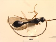
 Fig 1. Male, dorsal view |
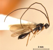 Fig 2. Male, profile |
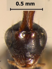 Fig 3. Head, front view |
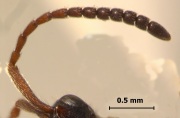 Fig 4. Antenna (female) |
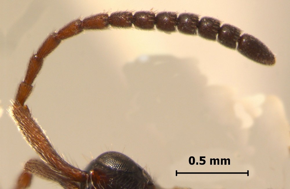
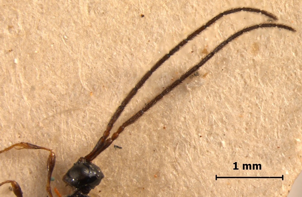
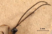 Fig 5. Antenna (male) |
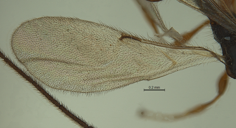
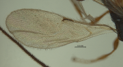 Fig 6. Forewing |
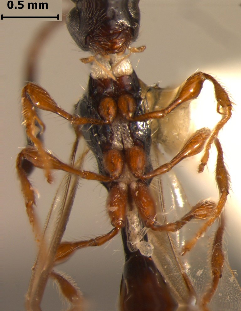
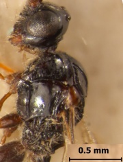 Fig 7. Mesosoma |
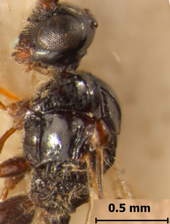
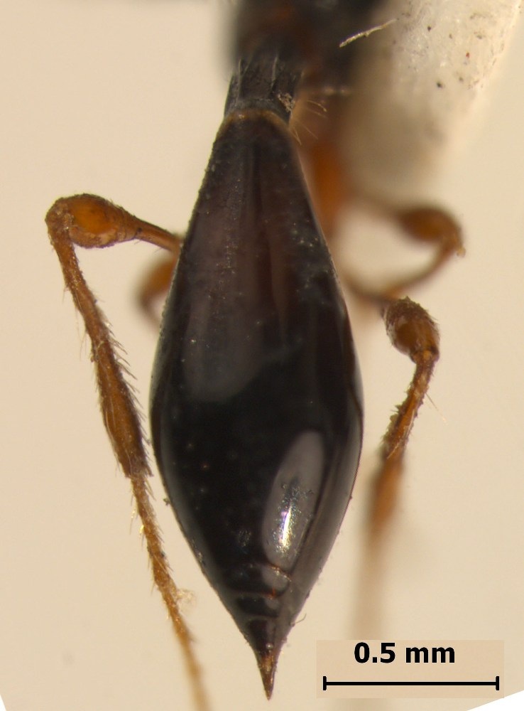
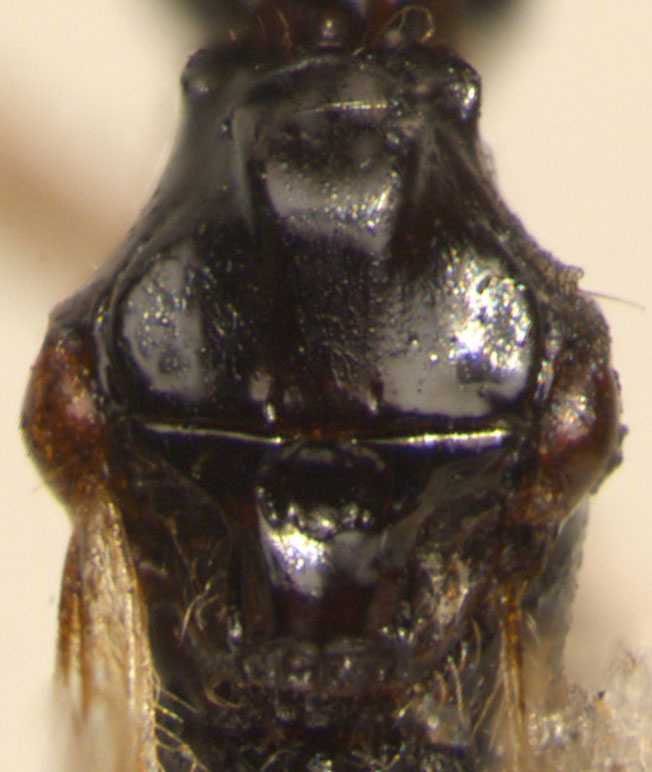
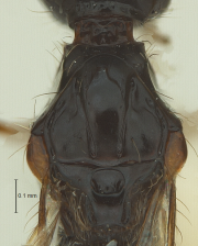 Fig 8. Anterior mesosoma |
 Fig 9. Metasoma (female) |
|
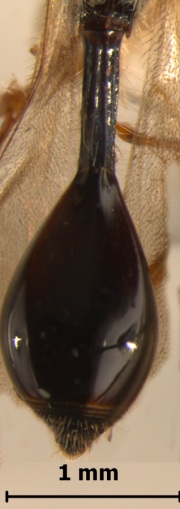 Fig 10. Metasoma (male) |
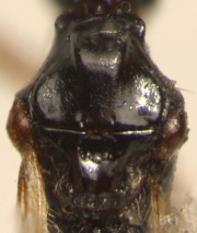 Fig 11. Notauli incomplete |
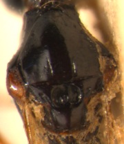 Fig 12. Notauli incomplete |
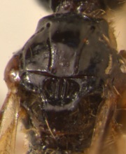 Fig 13. Notauli complete |
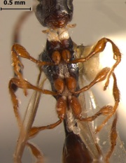 Fig 14. Foamy structures |
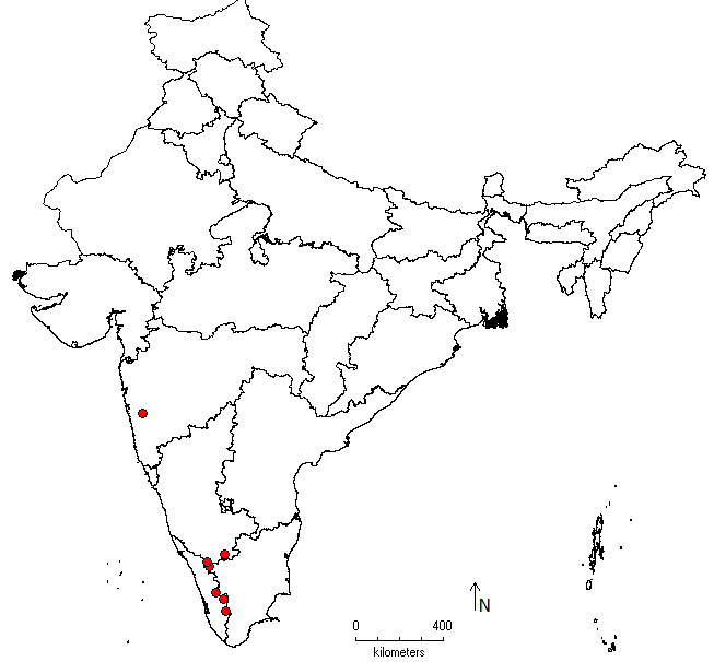
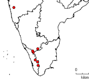 Fig 15. Distribution map |
|
Nomenclature
Paramesius Westwood, 1832. 129. Type species: Paramesius rufipes Westwood.
Aparamesius Kieffer, 1913: 436. Synonymized by Masner in Krombein and Burks, 1967.
Diagnosis
Medium to large, robust forms (usually 2-4mm). Head and body usually black or deep brown, with appendages much lighter in colour, body smooth and shining, lacking micropilosity, but with sparse long semierect hairs, also with foam-like structures on propleuron, intercoxal spaces and petiole (best visible ventrally). Hairy cushions on postgena, sides of pronotum and upper part of propleuron sparse.
Head subglobular. Antennal shelf often well developed when viewed laterally. Eyes large, often longer than temples (dorsal view). Mandibles normal, not beak-like. Malar sulcus not distinct. Antennae in both sexes 13-segmented. Female antennae without a distinct clava, terminal segments gradually enlarged, A13 longest and largest of clavomeres. Male antennae filiform, as long as or longer than body, segments elongate, narrow and cylindrical. A3 small, distinctly shorter than A4, subequal to A2. A4 onwards elongate, modified, slightly emarginate, often carinate. Pilosity on antennae short, not as long as width of A2. Occipital flange step-like.
Cervix distinct. Pronotum with shoulders often angulate. Propleuron variably sculptured. Mesoscutum with notauli absent or variably indicated, either as a linear furrow or much reduced to small oval pits. Anterior parapsidal lines often indicated. Humeral sulcus usually well developed, at times crenulate. Suprahumeral sulcus present occasionally. Anterior scutellar pit often monofoveate, rarely bifoveate, fovea at times with complete or incomplete longitudinal keels. Lateral and posterior scutellar pits sometimes present. Sternaulus usually indicated. Dorsellum with three keels medially. Propodeum sometimes elongate, with longitudinal keels, median keel produced, anteriorly raised and spine-like. Area between median keel and lateral ones (plicae) bare. Metapleuron with longitudinal striae, much concealed by fine dense hairs. Nucha indicated in some cases.
Wings often well developed, distal margin of forewings entire and rounded, not notched. Venation extending to half of forewing length. Costal cell narrow and tubular. Marginal vein elongate, many times length of stigmal vein, latter much reduced, post marginal vein absent, basal vein nebulous, present as a colouration, other veins lacking. A few species have a white hairless transverse streak on forewing medially. Hindwings with submarginal vein complete. Marginal cilia of forewings short, hardly reaching one-eighth of wing width.
Petiole elongate, tube-like, more than 2x longer than wide, usually with longitudinal striae and pilosity. Metasoma elongate. Anterior margin of T2/ large tergite not elevated above level of petiole, but on same plane, without a median furrow or emargination at base, rarely with two hairy depressions anterolaterally. Basal tergites visible as rings under large tergite.
Geographic Distribution
See Fig. 15 for Indian distribution of the genus.
Species known from India
1. Paramesius deccanus Sharma
2. Paramesius incompletus Kieffer
3. Paramesius malabarensis Rajmohana & Narendran
4. Paramesius monticola (Kieffer)
5. Paramesius pleuralis Kieffer
Remarks
Members of this genus can be diagnosed by their robust body, raised antennal shelf, normal mandibles, 13-segmented antennae in both sexes, in females antennae lacking an abrupt clava and A13 larger than A12, variably indicated notauli, anterior scutellar pit monofoveate, forewing large and wide, with reduced marginal cilia and anterior margin of T2 being concave.
This genus can be easily separated from Spilomicrus by the absence of foamy structures, elongate marginal vein, A13 in females being larger than A12, A3 distinctly shorter than A2 in males and T2 being articulated with petiole at the same level.
References
Kieffer. J. J. 1913. Serphides des Îles Philippines. Insecta, 3: 253-462.
Krombein K. V., and Burks, B. D. 1967. Hymenoptera of America north of Mexico. Synoptic Catalog. United States Department of Agriculture Monograph 2(2nd suppl.): 1-584. Washington.
Rajmohana, K and Bijoy, C., 2012. A checklist of Diapriidae and Proctotrupidae (Hymenoptera: Insecta) from India.
Westwood, J. O. 1832. Descriptions of several new British forms amongst the parasitic hymenopterous insects. London & Edinburgh Philosophical Magazine and Journal of Science 1: 127-129.
|